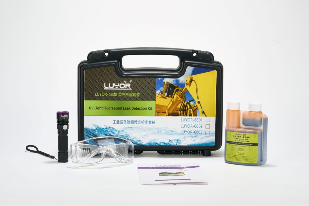
一套荧光检漏套装解决工业设备泄漏
LUYOR-6801荧光检漏仪是美国路阳公司生产的荧光检漏仪套装,适用于检查小到中等规模的油基液体系...
2024-03-28

作者:时间:2019-10-11 16:52浏览9027 次
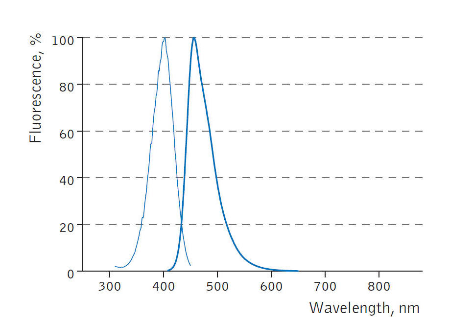
经常有客户咨询BFP(Blue Fluorescent Protein)的激发波长和发射波长,现在将BFP的激发波长和发射波长的光谱图提供给大家参考。BFP的激发波长在400nm左右,发射波长在450nm左右。
经常有客户咨询BFP(Blue Fluorescent Protein)的激发波长和发射波长,现在将BFP的激发波长和发射波长的光谱图提供给大家参考。
蓝色荧光蛋白BFP的激发光波长在400nm左右,发射光波长在450nm左右。

蓝色荧光蛋白的激发和发射光谱图
如果要观察BFP蓝色荧光蛋白(Blue Fluorescent Protein)的表达,美国路阳生产的便携式荧光蛋白激发光源可以选择LUYOR-3260VI和LUYOR-3415VX系列双波长荧光蛋白激发光源。蓝色荧光蛋白采用美国路阳紫光激发,佩戴LUV-20A绿色观察眼镜观察,如希望提供更多详细信息,可直接联系上海路阳生物技术有限公司的销售客服。
蓝色荧光蛋白Blue/UV Proteins
| 荧光蛋白名称 | 激发光波长 | 发射光波长 |
|---|---|---|
| TagBFP | 402 | 457 |
| mTagBFP2 | 399 | 454 |
| Azurite | 383 | 450 |
| EBFP2 | 383 | 448 |
| mKalama1 | 385 | 456 |
| Sirius | 355 | 424 |
| Sapphire | 399 | 511 |
| T-Sapphire | 399 | 511 |

Blue Fluorescent Proteins
Fluorescent proteins emitting in the blue region of the visible light spectrum (approximately 440 to 470 nanometers) were first obtained from site-directed mutagenesis efforts targeted at the tyrosine amino acid residue at position 66 in the GFP chromophore (see Figure 2). Conversion of this residue to histidine (Y66H) produces a blue fluorescent protein (BFP) that exhibits a broad absorption band in the ultraviolet centered close to 380 nanometers and an emission maximum at 448 nanometers. The original protein exhibited only about 15 to 20 percent of the parent GFP brightness value due to a low quantum yield and required additional secondary mutations to increase its folding efficiency and expression levels. Subsequent investigations and several additional mutations led to an enhanced BFP version that is still only 25 percent as bright as the enhanced green variant and displays limited photostability compared to many other fluorescent proteins.
The primary motivation for developing blue fluorescent proteins in the mid-to-late 1990s was the keen interest in creating matched pairs for fluorescence resonance energy transfer (FRET) experiments and multicolor labeling. Because the spectral characteristics (fluorescence emission profile) of BFP are readily distinguishable from EGFP, this protein combination was one of the first utilized for multicolor imaging. Blue fluorescent protein also has the distinction of being incorporated into the first genetically encoded biosensor along with an enhanced GFP variant to demonstrate FRET through linkage of the two fluorescent proteins via an intervening protease-sensitive spacer. The broad emission peak of BFP overlaps to a significant extent with the excitation spectrum of red-shifted GFP variants to yield a Förster distance of 4.1, a reasonable value for measuring FRET. Blue fluorescent protein has also been coupled with several GFP derivatives into biosensors designed to monitor transcription factor dimerization, calcium, and apoptosis.

Aside from the limited brightness levels and rapid photobleaching, blue fluorescent proteins also suffer from the fact that they must be excited with ultraviolet light, which is phototoxic to mammalian cells, even in limited doses. Furthermore, working in this spectral region is often hampered by autofluorescence and high absorption levels by cells and tissues, as well as light scattering. Microscopes operating in the ultraviolet also require specialized light sources, optics, and filter combinations that further complicate imaging. For all of the reasons listed above, the quest for more efficient blue fluorescent proteins has only been pursued by a few research groups. Investigations of mutagenesis using non-natural amino acids positioned in and around the chromophore have led to several blue-shifted "artificial" fluorescent protein variants that may find utility in several biological and photophysical applications.
Illustrated in Figure 2 are the chromophore structures for color variants of green fluorescent protein. In all cases, the first step in maturation of the chromophore is a series of torsional adjustments that relocate the carboxyl carbon of the amino acid at position 65 so that it is in close proximity to the amino nitrogen of the glycine residue at position 67 (Gly67) in the polypeptide backbone. Nucleophilic attack by this carbon atom on the amide nitrogen of glycine, followed by dehydration, results in formation of an imidazolin-5-one heterocyclic ring system. Fluorescence occurs when oxidation of the aromatic amino acid (position 66) carbon bond by molecular oxygen extends electron conjugation of the imidazoline ring system to include the aromatic substituent. The cyan, green, and yellow fluorescent protein variant chromophores are discussed in greater detail in the following sections.
 关注我们
关注我们