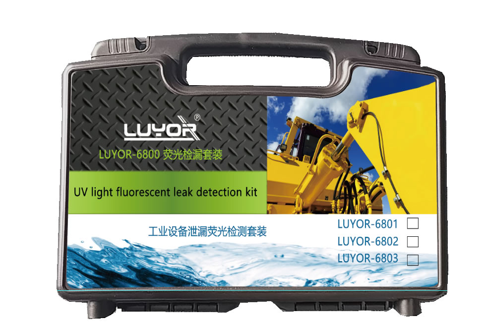
一套荧光检漏套装解决工业设备泄漏
LUYOR-6801荧光检漏仪是美国路阳公司生产的荧光检漏仪套装,适用于检查小到中等规模的油基液体系...
2024-03-28

作者:时间:2019-10-13 18:31浏览5887 次
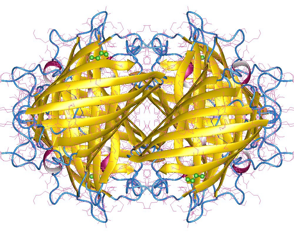
Venus荧光蛋白的激发光波长是515nm,发射光波长是529nm左右。Venus荧光蛋白属于黄色荧光蛋白的一种,所以,Venus荧光蛋白的发射光光谱图可参考黄色荧光蛋白的光谱图。Venus Fluorescence Protein is a basic (constitutively fluorescent) yellow fluorescent protein published in 2002, derived from Aequorea victoria. It is reported to be a rapidly-maturing weak dimer with moderate acid sensitivity.
Venus荧光蛋白的激发光波长是515nm,发射光波长是529nm左右。Venus荧光蛋白属于黄色荧光蛋白的一种,所以,Venus荧光蛋白的发射光光谱图可参考黄色荧光蛋白的光谱图。

Venus Fluorescence Protein
Venus is a basic (constitutively fluorescent) yellow fluorescent protein published in 2002, derived from Aequorea victoria. It is reported to be a rapidly-maturing weak dimer with moderate acid sensitivity.
Yellow Fluorescence Proteins
| Protein | λex | λem | Notes |
|---|---|---|---|
| EYFP | 513 | 527 | |
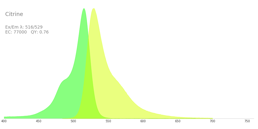
| Citrine | 516 | 529 | EYFP derivative; Less Cl, pH sensitive |
| Venus | 515 | 528 | EYFP derivative; 30-fold brighter at 37° C |
| SYFP2 | 515 | 527 | improved Venus |
| TagYFP | 508 | 524 |
如果要观察Venus黄色荧光蛋白(Venus Fluorescent Protein)的表达,美国路阳生产的便携式荧光蛋白激发光源可以选择LUYOR-3260CY和LUYOR-3415C(X)系列双波长荧光蛋白激发光源。黄色荧光蛋白采用青色光源激发,佩戴LUV-40A桔色观察眼镜观察,能够分选愈伤组织、菌落、植株、种子等黄色荧光蛋白表达阳性的目标,是转基因筛选的良好工具。如希望提供更多详细信息,可直接联系上海路阳生物技术有限公司的销售客服。


上图为LUV-40A黄色荧光蛋白观察眼镜

上图为LUV-550A 黄色荧光蛋白拍照滤镜
Yellow fluorescent protein (YFP) is a mutant variant of GFP. In this case, a mutation was introduced after the discovery that threonine residue was present near the chromophore in GFP.
To bring stability to the excited dipole moment of chromophore, this threonine residue was mutated. This led to shift by 20 nm in the excitation and emission wavelength of the fluorescent protein, leading to longer wavelengths.
YFP was further improved that led to development of enhanced yellow fluorescent protein or eYFP. This is one of the brightest fluorescent proteins and is widely used for different imaging purposes. GFP and YFP are used in several cases to perform dual imaging, which is a procedure where two molecules are tagged to two different fluorescent proteins. Consequently, they can be imaged together in order to visualize and appraise their inter-dynamics.
 关注我们
关注我们